OCR Physics
Gamma imaging
6.5.2 Diagnostic methods in medicine (Gamma)
6.5.2-1 the gamma camera
It is a gamma detector for a γ source injected to the patient.
γ is a suitable source, because:
- Least ionising, so least harmful;
- Most penetrative, so can be detected easily outside the body.
Radioactive Isotopes (Radioisotope) for γ camera should have short half-life, because:
- High activity of the source to form an image from small amount of radiation;
- Patient is exposed to radiation in a shorter period.

Radioisotopes:
- Fluorine-18: used in PET scans, has a short half-life and is produced at the hospital.
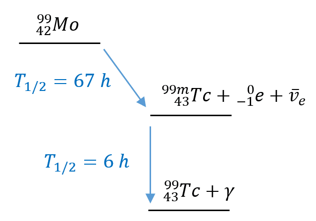
- Technetium-99m (Tc-99m): is daughter nucleus of
 decay of molybdenum-99 (Mo-99). It is used to check function of major organs, e.g. brain, lung, kidney, liver, heart. The “m” in Tc-99m means metastable which means Tc is unstable and will decay soon with γ of energy of exactly 140 keV and half-life of 6 hours.
Medical Tracers (radiopharmaceuticals):
Radioisotopes are bonded with chemicals which can be absorbed by our organs. This combination is called “Medical Tracers” or radiopharmaceuticals.
Tc-99m + sodium and oxygen = ![]() -> targets brain.
-> targets brain.
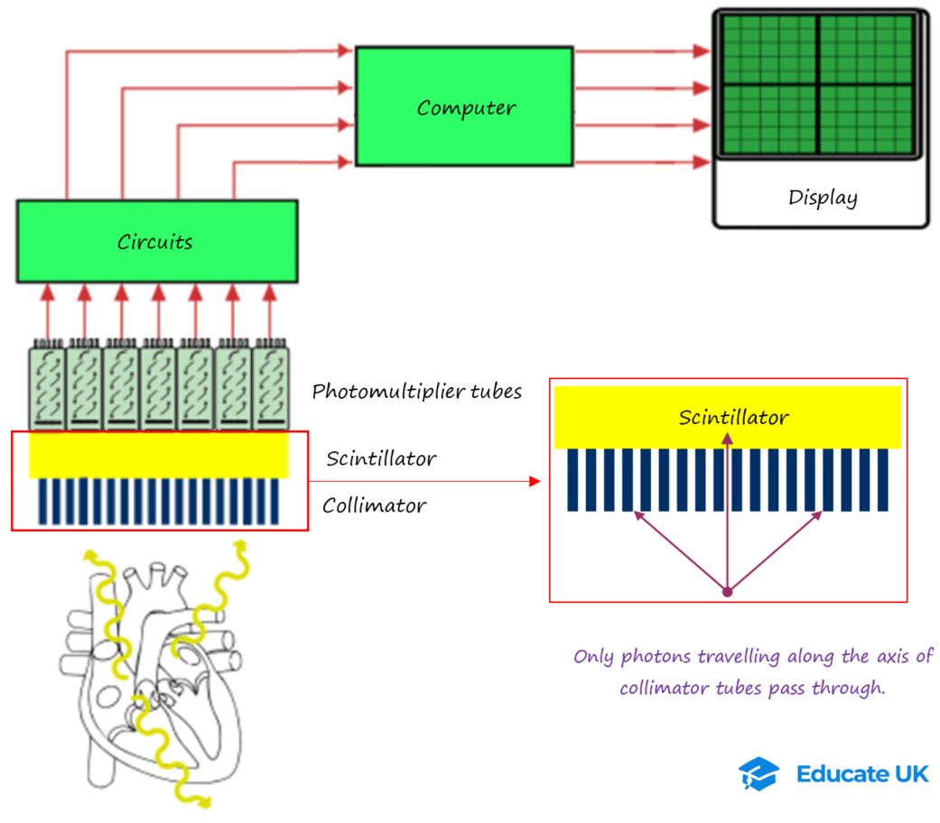
The γ produced by Tc-99m is detected by a gamma camera.
Gamma Camera:
X-ray shows the anatomy of an organ…
But Gamma camera shows if an organ is functioning properly!
Gamma camera image source: Link and Link. (Image modified.)
Gamma photons emitted from the patient, first enter the collimator made of lead tubes. Only those pass through that are parallel to tubes of collimator. Rest are absorbed by the tubes.
Then the photons get to the scintillator made from sodium iodine. Here each photon of γ produces thousands of visible light photons.
Probability of γ photon reaching the scintillator is 1/10.
The visible light goes into the photomultiplier tubes.
The photomultiplier converts each visible photon to electrical pulse (Potential Difference). The PD is converted to image with a sophisticated software. That’s all you need to know now!
The photomultiplier
It is a vacuum glass tube which increases the number of electrons.
Visible light photon passes through photocathode and releases a photoelectron (photoelectric effect).
The photoelectron is attracted by the first electrode (dynode) with a PD of +100 V.
Due to impact of the photoelectron with +100 V dynode, four secondary electrons are released.
These four are attracted to the second dynode with PD of +200 V.
And each produces four more electrons and so on…
After 10 dynodes there will be 410 electrons.
At the end the anode collects all the electrons which produces a tiny voltage pulse through a resistor (electron read-out shown below):
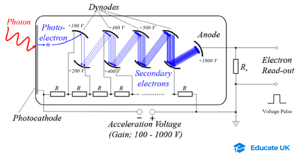
Image source: link (Modified).
Figure below shows the left kidney is infected as it has taken less Tc-99m and hence is not as active as the right kidney.
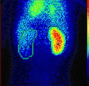
Image source: Link.
When describing the workings of a gamma camera always say gamma photon or visible light photon; not gamma ray or just visible light!!
PET Scans:
Positron emission tomography
Just like CAT scans, PET also produces 3D images with high details, but in PET γ is used.
Fluorine-18 (![]() ) is used in PET scans.
) is used in PET scans.
Decay equation:

The positron emitted from F-18 decay, annihilates with an electron within patient. The pair of photons from annihilation are detected by the machine.
Note: decay of F-18 itself produces a γ photon, but this is not detected by the PET scanner.
F-18 has a half-life of about 110 minutes. Because of short half-life it has to be made in the hospital by a particle accelerator.
In particle accelerator a proton is collided with Oxygen-18 (O-18 constitutes 20% of natural oxygen), and ![]() is produced + neutron:
is produced + neutron:

To target body organs Fluorine-18 is usually bonded with fluorodeoxyglucose (FDG).
In FDG, F-18 replaces an Oxygen atom.
Our body sees FDG as glucose, hence it accumulates in organs with high respiration.
F-18 also is bonded with carbon monoxide (CO) made with carbon-11 isotope, which is a positron emitter with half-life of 20 minutes.
CO bonds well with haemoglobin molecule in red blood cell.
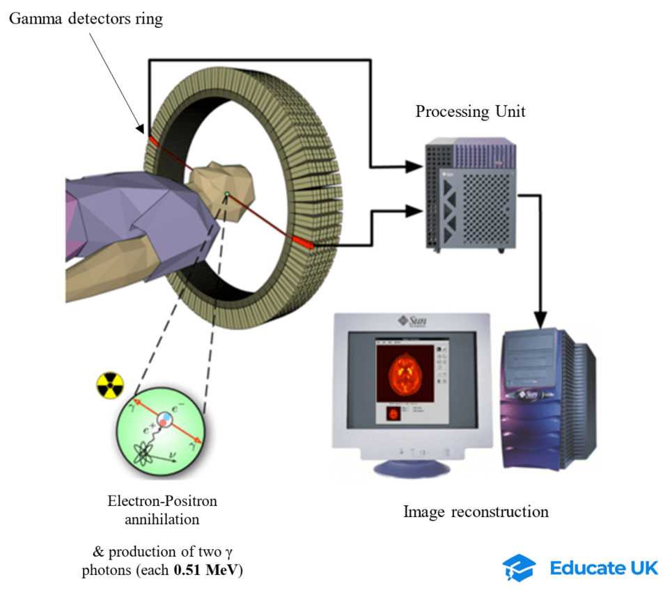
PET scanner:
Two photons from electron-positron annihilation is detected by the ring of gamma detectors.
Each photon passes through the scintillator (made of sodium iodine) of the detector, and then the photomultiplier which produces a pulse of voltage.
The two photons move in diametrically opposite direction (thanks to conservation of momentum!). Then the computer can determine the location of annihilation by measuring the difference in time of photons received by the ring of detectors, and using speed of EM waves (3 × 10⁸ m/s).
Advantages of PET scan:
- Non-invasive diagnosis;
- Used in cancer diagnosis, planning heart surgery, examine brain function
- Identifying start of brain problems e.g. Alzheimer disease.
Disadvantages:
- Very expensive, only available at large hospitals;
- Only patients with serious problems are taken for PET scans.
.
Revise and Get Paid!
If you like taking summary notes of lessons and solving past papers, see the Join Us page!