OCR Physics
Ultrasound
6.5.3 Ultrasound
Human can hear sound in frequency range of 20 Hz to 20 kHz.
Ultrasound is any frequency above 20 kHz.
Advantages of ultrasound:
- Non-invasive diagnosis;
- Non-ionising, it’s just sound; no radiation!
- Quick
Frequency of ultrasound for medical imaging ranges 1 – 15 MHz.
Ultrasound travelling through body can be:
- Reflected;
- Refracted: when traveling between different tissues;
- Diffracted: when going through gaps.
Ultrasound transducer: device that produces and receives ultrasound with help of Piezoelectric Effect…
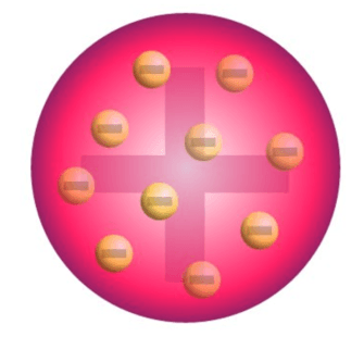
Piezoelectric effect:
Some crystals, e.g. quartz, produce an e.m.f. when deformed mechanically.
Also if an e.m.f. is applied to them, they deform!
Max strain of quartz is 0.1%.
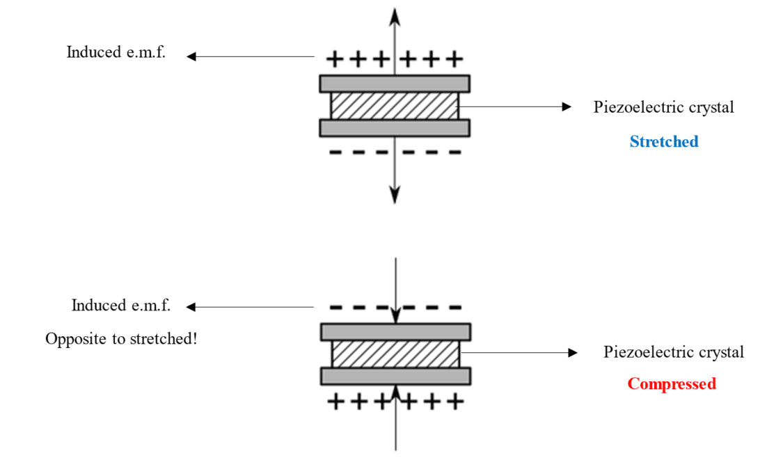
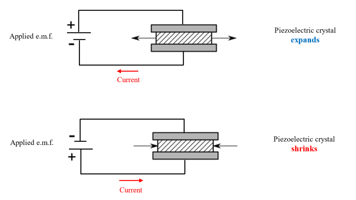
Image source: Link.
Ultrasound transducer:
To produce ultrasounds an Alternating P.D. of frequency 5 MHz is applied to a piezoelectric crystal.
This frequency is the same as the natural frequency of the crystal, which causes resonance. And vibrations in turn produce ultrasound of frequency 5 MHz.
The reflected ultrasound from body tissue is received by the same transducer which causes Alternating e.m.f. in the crystal.
Today the piezoelectric used is either lead zirconate, or polyvinylidene, instead of quartz.
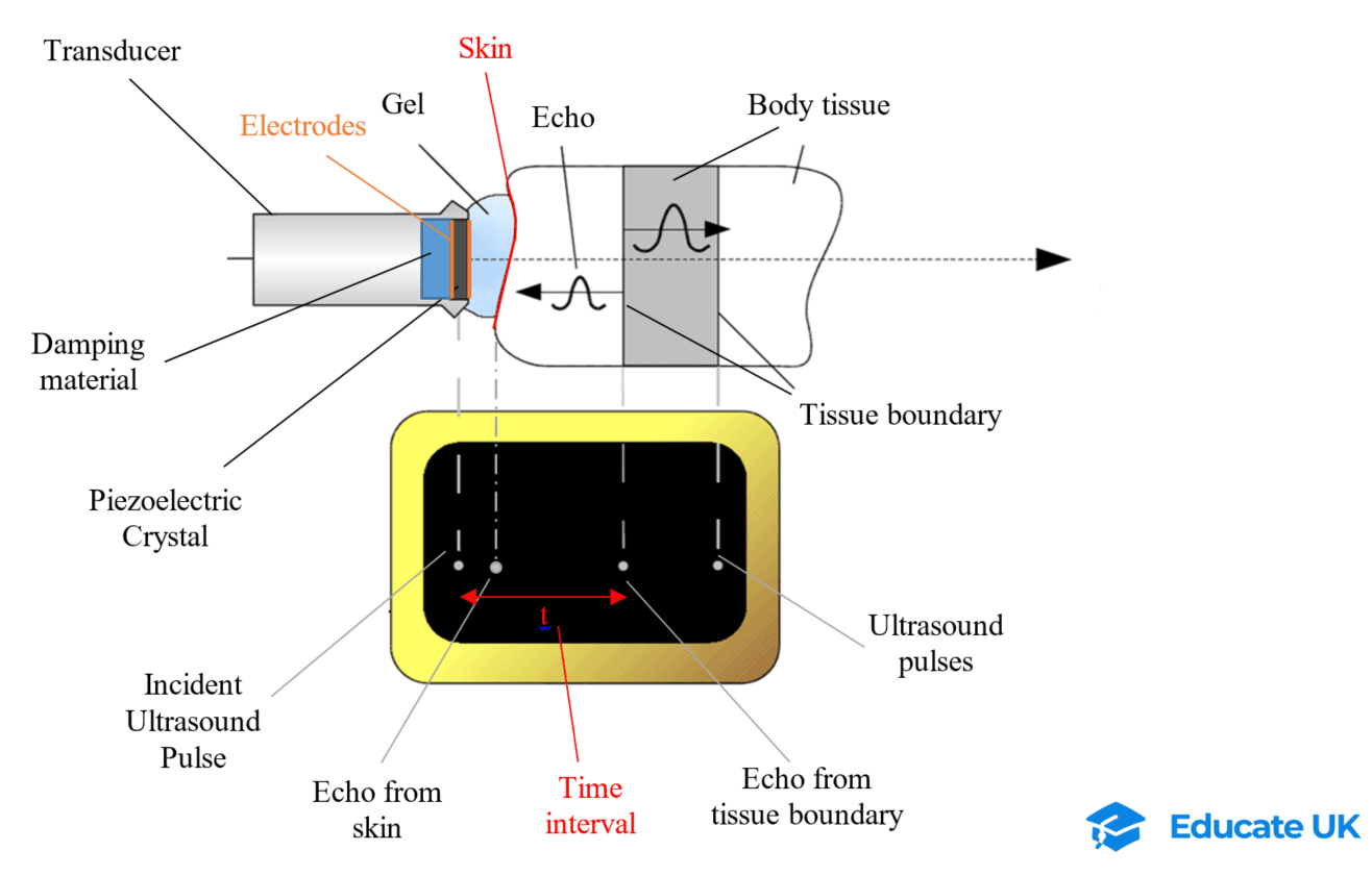
Image source: Link.
6.5.3-1 Ultrasound Scans:
A-Scan:
Simplest form of ultrasound.
A single transducer records the pulses and their reflection along a straight line.
It can be used to measure thickness of a bone, or distance between the retina and lens in an eye.
As the ultrasound goes through body tissues, some of it is reflected and some transmitted. As a result, the reflected sound will have less energy than the original wave because some of it is transmitted and passes through rather than reflect!
The time taken from when the original sound is released to when its reflection is received is called the time interval.
If the average speed of the ultrasound in the body tissue is known, using the time interval, distance between two reflections in the body is calculated.
Example:
If the average speed of ultrasound in the eye is 1550 m/s, and the time interval of an A-Scan for reflections at the front and back of the eye lens is 2 μs and 7 μs; calculate the thickness of the lens.
Total distance travelled by ultrasound in 5 μs:
Distance = vt = 1550 × 5 × 10-6 = 7.75 × 10⁻³ m.
This distance is for ultrasound to travel from transducer and back, so thickness of eye lens:
(7.75 × 10⁻³) / 2 = 3.875 × 10⁻³ m.
B-Scans
Most commonly used type.
Also known as 2D scan.
It is combination of multiple A-scans.
As the transducer is moved on the skin and its angle is changed, the transducer records the reflection due to boundary between different body tissues.
Each reflection produces a dot on a digital screen.
The brightness of the dots depends on the intensity of the reflection.
The combination of all dots on the screen forms a 2D image.
The B in B-scan comes from Brightness!
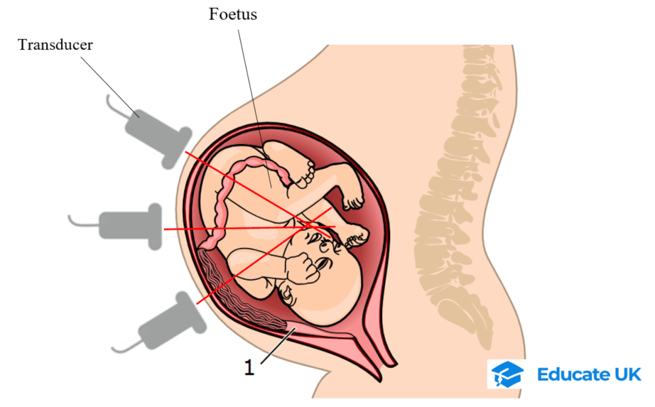
Image source: Link. Image modified.
6.5.3.-2 Acoustic impedance
Ultrasound wave reaching the boundary between two tissues, will either reflect or refract.
The proportion of reflected wave depends on the acoustic impedance (z) of both tissues.
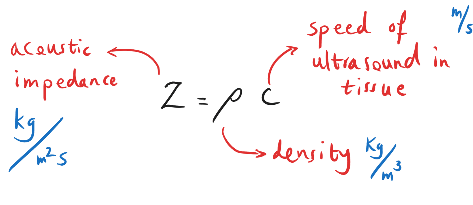
Intensity reflection coefficient
The intensity of reflected ultrasound wave at a boundary, depends on acoustic impedance of the two material at the boundary.
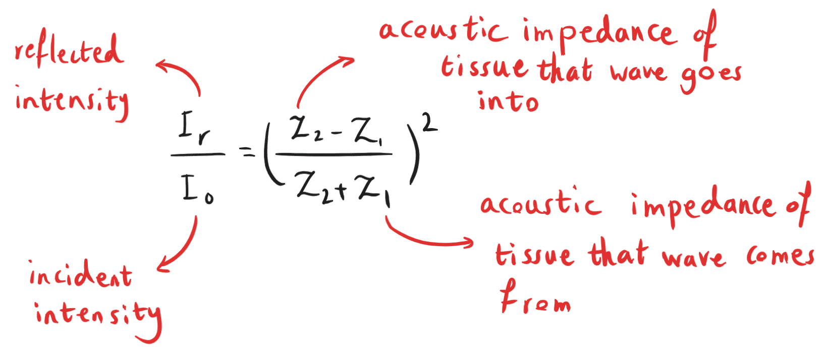
The ratio ![]() is called intensity reflection coefficient!
is called intensity reflection coefficient!
The higher the difference between z1 and z2, the bigger the reflection intensity.
As the fraction is squared, it does not matter which z is for which material!
Example 1:
An ultrasound beam is incident at 90o angle at a muscle-bone boundary. If acoustic impedance of muscle and bone are 1.69 and 7.6 respectively, find the intensity reflection coefficient at this boundary.
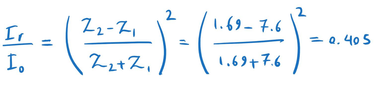
This means 40.5 % of the ultrasound is reflected, the rest is refracted through the next medium.
Acoustic matching
By placing an ultrasound transducer on the skin some air is trapped between the transducer and the skin. The causes almost all (99.9%) of the ultrasound to reflect from the skin and none to get to the body tissues.
To solve this, a coupling gel is smeared on the skin first, which eliminates the air bubbles.
The acoustic impedance of the coupling gel is very similar to that of the skin.
This is called acoustic (or impedance) matching.
6.5.3-3 Doppler Ultrasound
Doppler Effect: frequency of wave changes if the wave source moves towards or away from us.
This also happens if wave is reflected from a moving object.
Reflection of ultrasound from red blood cells, is used to examine the blood flow and find out if there are any blood clots (thrombosis) or fat deposits (atheroma) in the main veins and arteries.

Image source: Link.
Reflected ultrasound:
- From stationarytissue: same frequency and wavelength;
- From blood cells moving away from transducer: decrease in frequency;
- From blood cells moving towards the transducer: increase in frequency.
The change in frequency depends on the speed of blood.
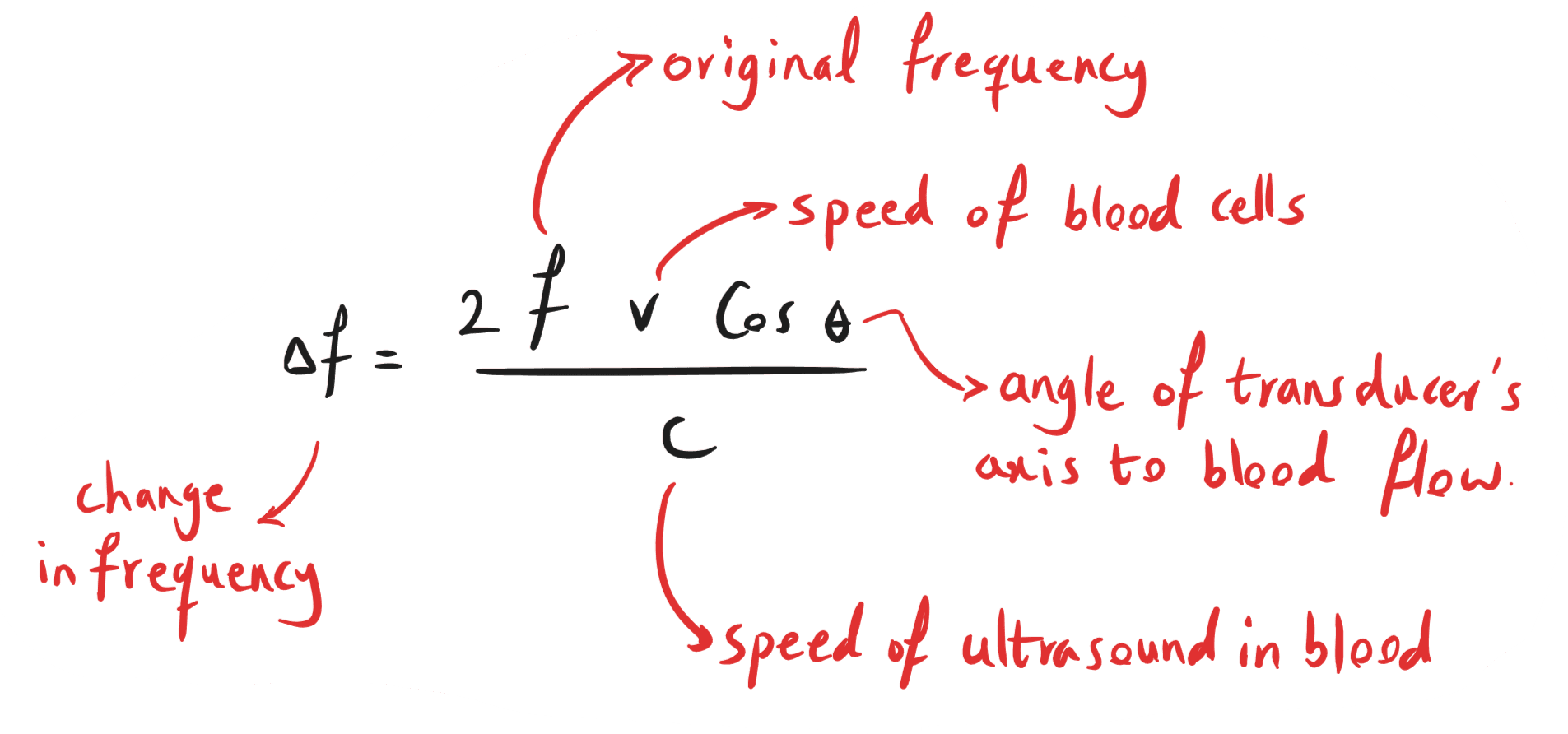
.
Revise and Get Paid!
If you like taking summary notes of lessons and solving past papers, see the Join Us page!