OCR Physics
X-Ray Imaging
6.5.1 X-Ray
X-ray wavelength range: 10-8 to 10-13 m.
X-ray can kill living cells. This is used in cancer treatment.
6.5.1-1 X-Ray tube:

(Image modified.)
The filament (cathode) which is connected to a low voltage, produces electrons (thermionic emission).
Electrons are attracted to the positive target metal (anode).
A high voltage source provides the PD between the anode and cathode.
The fast moving electrons remove an electron close to the nucleus of the target metal (anode) and create a gap.
The gap is filled by electrons from higher energy levels of the atom and as a result X-ray photons are produced.
One electron produces one photon!
6.5.1-2 X-ray attenuation
Bones absorb more X-ray than soft tissue. Soft tissue scatters the rays. Both of these cause reduction in the intensity of X-ray, this reduction is called attenuation.
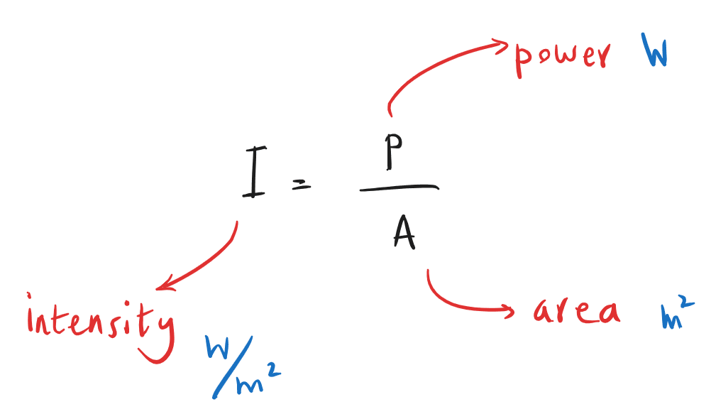
Intensity:
Attenuation mechanisms:
| Simple Scatter
X-ray with photon energy 1-20 keV.
Photon does not have enough energy to remove an electron, so the photon bounces off the electron (it is scattered) without change in energy of the photon. |
| Photo electric effect
X-ray photons with energy less than 100 keV. The photon is absorbed by an electron. The electron escapes the atom. This is dominant in hospital radiography as photons have 30-100 keV of energy. |
| Compton scattering
Photon energy range: 0.5-5 MeV. Photon ejects an electron and then is scattered with lower energy. In this process both energy and momentum are conserved. Used in killing cancerous cells. |
| Pair production
Photon energy more than or equal to 1.02 MeV. Photon interacts with nucleus. It disappears and an electron and positron are created. Used in killing cancerous cells. |


Images from: link.
Attenuation formula:
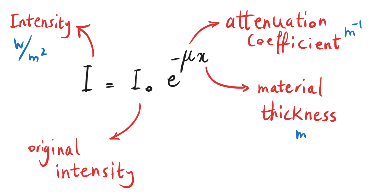
Bones have a bigger μ than muscles, because they absorb X-ray better!
The intensity of rays transmitted is recorded on an electronic sensor which produces a two dimensional image of the body.
6.5.1-3 Contrast medium
As soft tissue has low level of X-ray absorption, contrast material are used for a better image.
E.g. barium and iodine. They have large atomic number (z).
Attenuation coefficient is proportional to cube of z à ![]()
For soft tissue inside body average z = 7
Barium: z = 56 and iodine: z = 53
This means barium is 510 times and iodine is 430 times more absorbent of X-ray than soft tissue.
Iodine is added to liquids of body e.g. blood. The resulting X-ray image shows blockages of blood vessels or heart structure.
Barium sulfate is used to produce X-ray of digestive system. Patient drinks a liquid mixture called barium meal, just before X-ray image is taken.

Click on the link for the animation of barium swallow.
6.5.1-4 CAT Scans
Stands for computerised axial tomography.
A regular X-ray provides a two dimensional image. So it does not differentiate overlapping bones and tissues.
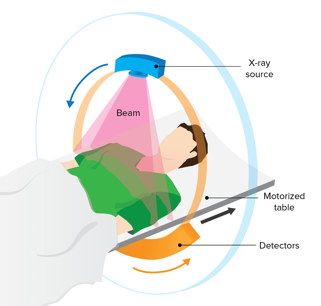
CAT scan takes X-ray images from different angles and combines them to a 3-D image.
The X-ray tube (source) produces a thin fan-shaped beam (pink beam shown below).
Both the tube and detectors make a 360o rotation around the patient.
Conventional X-Rays | CAT/CT Scans | |
| Imagery | 2D images | detailed 3D images |
| Detail | Less detailed; mainly shows bones and some organs | More detailed; shows shape, size of bones, blood vessels, and soft tissues or disorders |
| Speed | Quick and usually takes only a few minutes | Takes longer 15-30 minutes, exposure equivalent to several years of background radiation |
| Use | Commonly used for detecting fractures and infections. Cheaper | Often used for diagnosing conditions such as cancer, cardiovascular diseases, and internal injuries. Patient should remain still otherwise image is blurred |
.
Revise and Get Paid!
If you like taking summary notes of lessons and solving past papers, see the Join Us page!